Angew. Chem. Int. Ed. | Wu Dongdong/Bai Chen team explores ultra-thin peptide-based nanosheets.
In May 2024, Professor Wu Dongdong from the Biomedical Engineering College at Sichuan University and the MoMed Biotech team led by Bai Chen published a research paper titled "Multi-Responsive Peptide-Based Ultrathin Nanosheets Prepared by a Horizontal Monolayer Assembly" in Angew. Chem. Int. Ed. This work not only sets a new thickness record for peptide-based nanomaterials but also provides new ideas for functionalizing and intelligentizing 2D materials.
Material Introduction
Two-dimensional (2D) nanomaterials have attracted much attention due to their unique physical and chemical properties and their potential applications in multiple fields. In particular, 2D materials have shown great potential as drug delivery systems, biosensors, and tissue engineering scaffolds in the biomedical field. However, existing 2D materials often suffer from complex preparation and single functionality, which limits their effectiveness in practical applications. To overcome these limitations, the research team focused on peptide-based materials, utilizing their good biocompatibility and adjustable structure. Through an innovative covalent-physical manufacturing strategy, they successfully prepared ultra-thin nanosheets with a thickness of only 1 nanometer.
Research Testing
In this study, the research team used a layered covalent-physical manufacturing strategy to prepare peptide-based self-assembled nanosheets with a thickness of approximately 1 nanometer. By covalently alternating the assembly of the helical peptide E3 with azobenzene (AZO) structures, copolymers CoP(E3–AZO) were produced. These copolymers physically self-assembled into ultra-thin nanosheets, forming an unexpected two-dimensional horizontal monolayer arrangement. This unique monolayer arrangement made the thickness of the nanosheets equivalent to the cross-sectional diameter of a single linear copolymer. Typically, self-assembled molecules in nanosheets are stacked and arranged perpendicular to the nanosheet surface, and the thickness of the nanosheet is determined by the length of the self-assembled molecules, which is at least greater than 2 nm. Therefore, the parallel arrangement of self-assembled polymers reported here is a rare phenomenon in nanosheet formation.
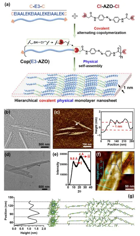
Figure 1. Formation and characterization of ultrathin nanosheets. (a) Schematic illustration of the synthesis of two-dimensional ultrathin nanosheets through covalent polymerization and physical self-assembly layer-by-layer process. (b) SEM image of the ultrathin nanosheets. (c) AFM image and height profile of the ultrathin nanosheets. (d) TEM image of the ultrathin nanosheets. (e) WAXS spectrum of the powder sample of the ultrathin nanosheets. (f) AFM image indicating the possible distance in the nanosheets corresponding to the two peak signals in the WAXS image. (g) Molecular cartoon illustrating regular height variation within a single nanosheet.
Using molecular dynamics simulations, it was found that a synergistic effect of various molecular interactions drove the self-assembly of CoP(E3-AZO) into nanosheets. Strong electrostatic interactions between molecules led to the aggregation of the copolymer, while π-π stacking within the nanosheets and anionic surfaces facilitated two-dimensional self-assembly. Additionally, hydrophobic effects and hydrogen bonding stabilized the two-dimensional polymer assembly.
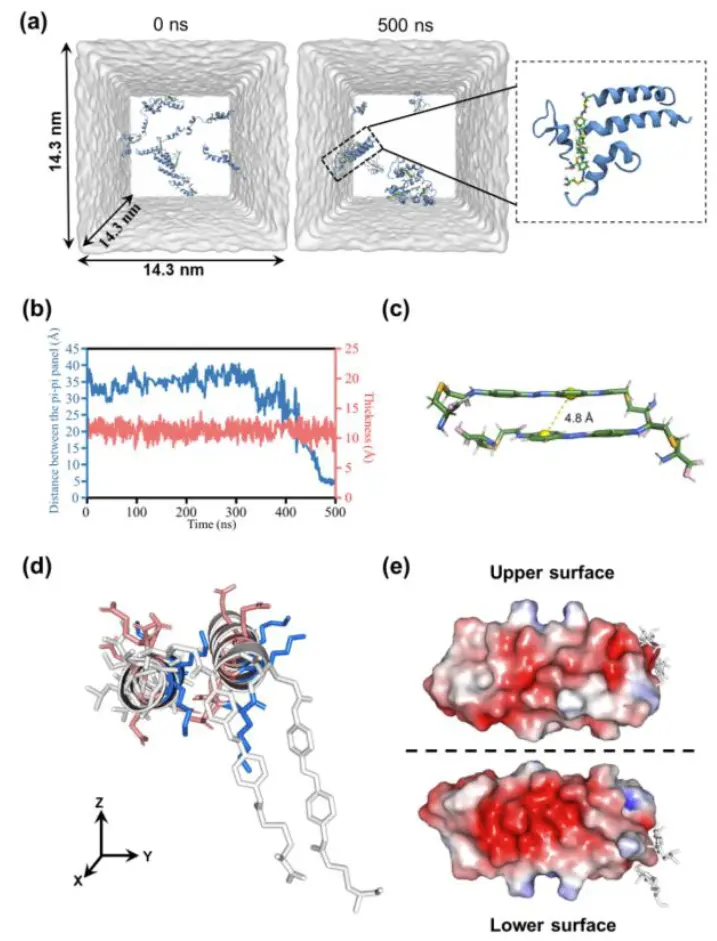
Figure 2. Molecular simulation of ultrathin nanosheets. (a) Molecular dynamics simulation of the self-assembly of copolymers of E3 and azobenzene. The model copolymer consists of two E3 segments and one azobenzene structure. The simulation shows the structure of the model copolymer in the initial state (0 ns) and the final state (500 ns). (b) Evolution of the distance between π-π panels and the thickness of the aggregated model polymer. (c) DFT optimized structures of two model polymers (with azobenzene); the distance between the π-π panels is 4.8 Å. (d) Configuration of parallel polymers. Lysine residues with positive charges are shown in blue, and glutamic acid residues with negative charges are shown in red. (e) Electrostatic potential on the upper and lower surfaces of the parallel polymer. Regions with positive charges are shown in blue, and regions with negative charges are shown in red.
In addition, the research team found that by using different methods such as light treatment, pH adjustment, addition of additives, and introduction of cosolvents, molecular interactions can be altered and the self-assembly of CoP(E3-AZO) can be regulated to produce diverse nanostructures.
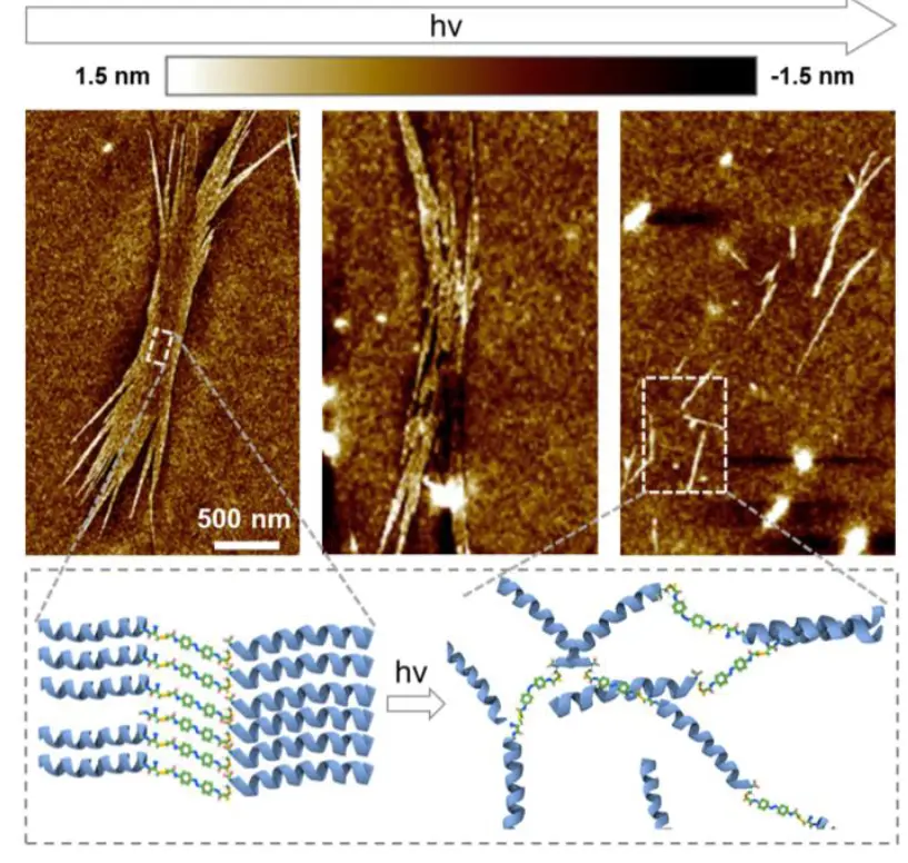
Figure 3. Photoresponse of ultrathin nanosheets.
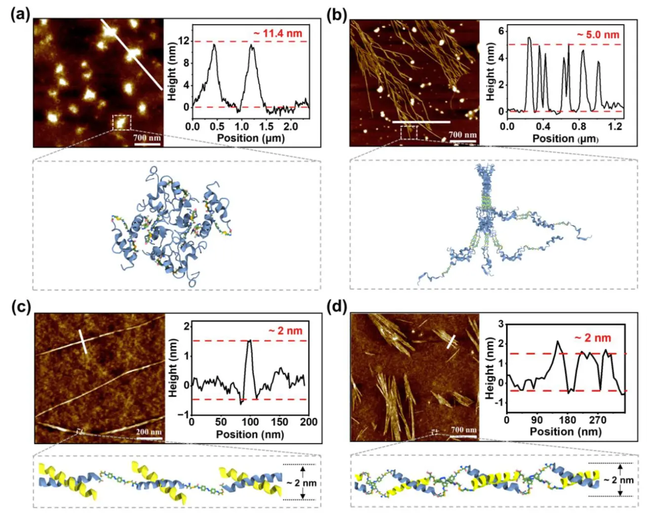
Figure 4. Self-assembly of CoP(E3-AZO) in different environments. AFM images of the self-assembly of CoP(E3-AZO) (a) at pH 4, (b) at pH 10, (c) with K3 molecules, and (d) with CoP(K3-AZO) copolymers. The plot on the right side of the AFM images shows the height distribution in the region indicated by the white line.

Figure 5. Evolution of the structure of CoP(E3-AZO) during self-assembly in 50 vol% CH3CN/H2O for 2, 10, 17, and 24 days. The plot below the AFM images shows the height profile in the region indicated by the white line.
It is noteworthy that these ultrathin nanosheets can selectively inhibit cancer cells at specific concentrations.
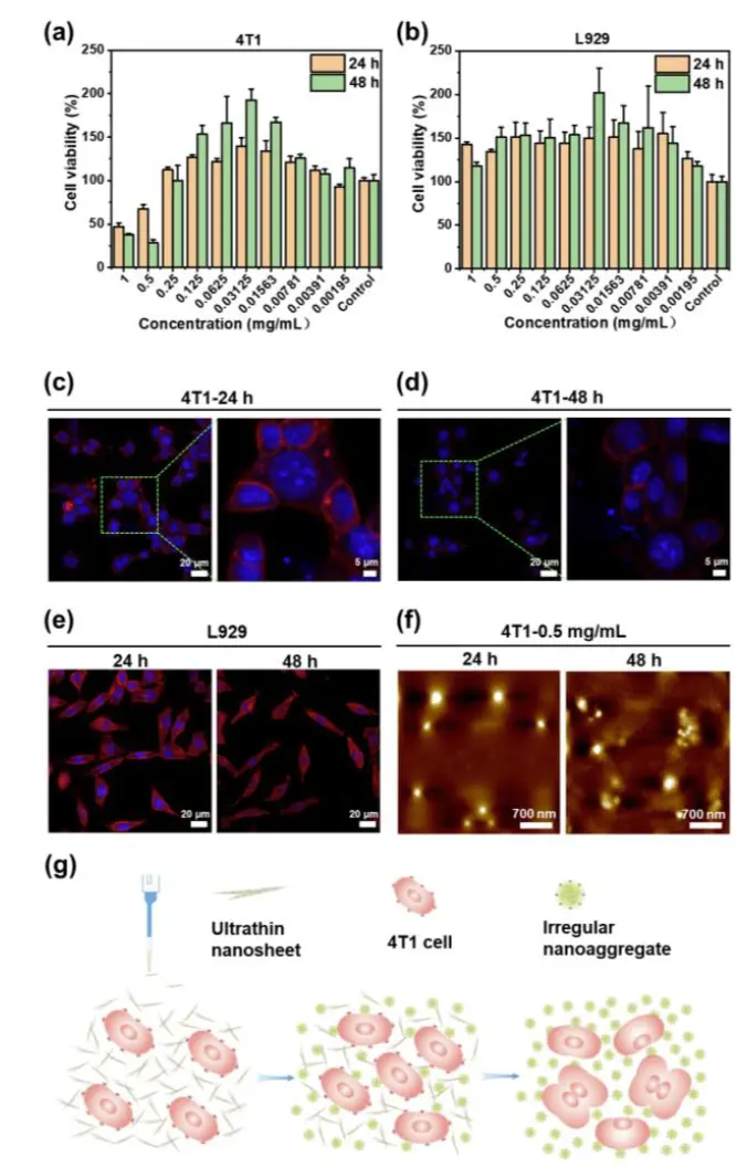
Figure 6. Anticancer activity of ultrathin nanosheets against 4T1 cells. (a) Relative cell viability (n=3) of 4T1 cells treated with ultrathin nanosheets at different concentrations. The error bars indicate the standard deviation. (b) Relative cell viability (n=3) of L929 cells treated with ultrathin nanosheets at different concentrations. The error bars indicate the standard deviation. (c) Morphology of 4T1 cells treated with 1 mg/mL ultrathin nanosheets for 24 hours. (d) Morphology of 4T1 cells treated with 1 mg/mL ultrathin nanosheets for 48 hours. (e) Morphology of L929 cells treated with 1 mg/mL ultrathin nanosheets for 24 and 48 hours. (f) AFM images of 4T1 cells incubated with 0.5 mg/mL ultrathin nanosheets for 24 and 48 hours. (g) Schematic showing the pH-responsive ultrathin nanosheets inhibiting the growth of 4T1 cells.
This study not only enriches the family of 2D nanomaterials but also provides a new platform for the development of biomedical materials. The intelligent responsive properties of ultrathin nanosheets endow them with enormous potential applications in sensing, catalysis, electrochemical applications, and beyond. Particularly in the biomedical field, they offer new strategies for cancer treatment.
Research Paper Original Link: https://doi.org/10.1002/anie.202405765
Hot News
-
2022-11-05
The Bai Chen Research Group Publishes Paper in the International Journal of Molecular Sciences
- 2022-12-20
- 2022-12-26
- 2026-02-25


